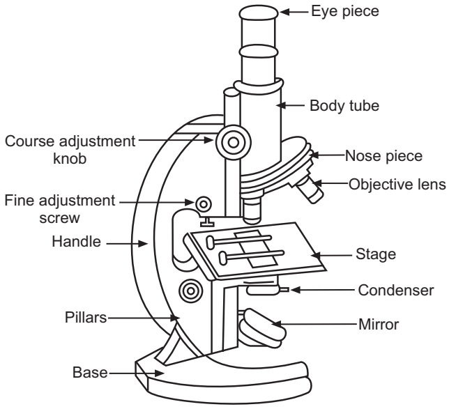
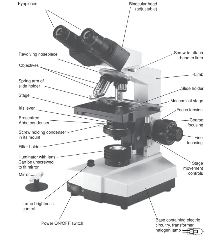
44 diagram of a microscope with label
Interactive Eukaryotic Cell Model - CELLS alive WebSecretory Vesicle: Cell secretions - e.g. hormones, neurotransmitters - are packaged in secretory vesicles at the Golgi apparatus.The secretory vesicles are then transported to the cell surface for release. Cell Membrane: Every cell is enclosed in a membrane, a double layer of phospholipids (lipid bilayer).The exposed heads of the bilayer are "hydrophilic" … Latest Breaking News, Headlines & Updates | National Post WebRead latest breaking news, updates, and headlines. Get information on latest national and international events & more.
PPIC Statewide Survey: Californians and Their Government Oct 26, 2022 · Key Findings. California voters have now received their mail ballots, and the November 8 general election has entered its final stage. Amid rising prices and economic uncertainty—as well as deep partisan divisions over social and political issues—Californians are processing a great deal of information to help them choose state constitutional officers and state legislators and to make ...
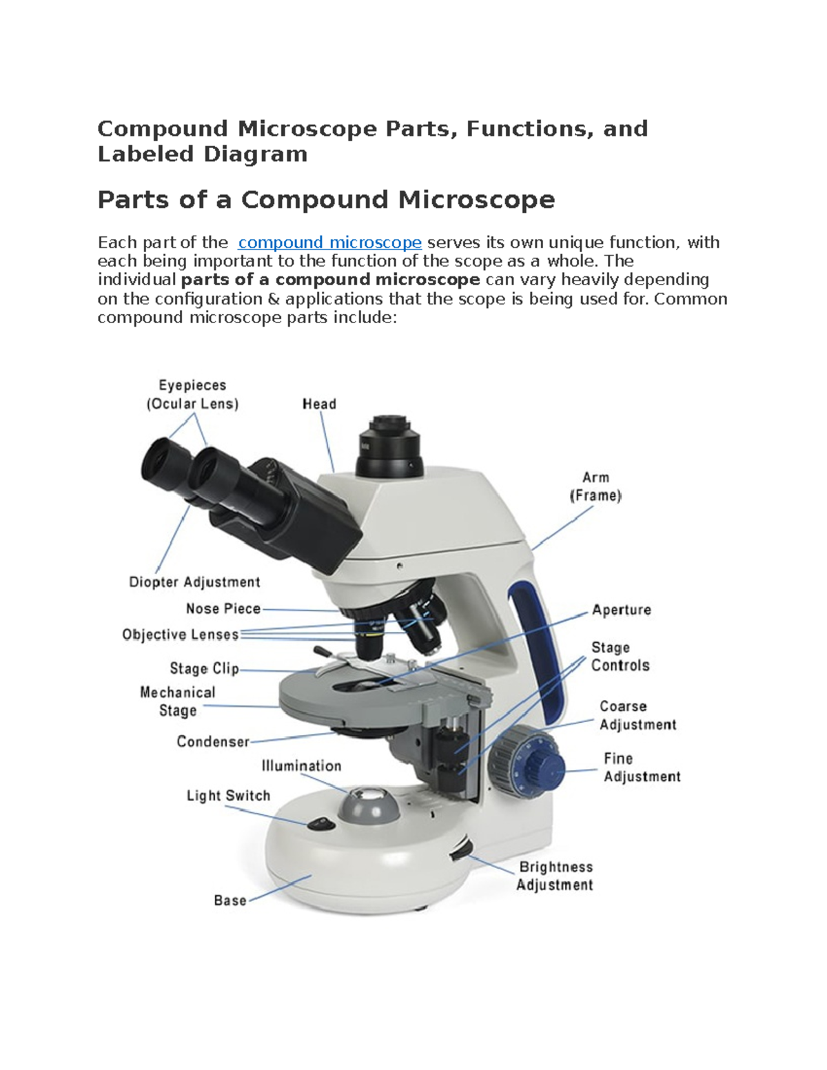
Diagram of a microscope with label
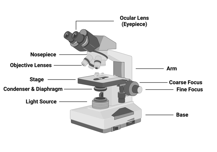
Compound Microscope Parts – Labeled Diagram and their … WebMajor structural parts of a compound microscope. There are three major structural parts of a compound microscope. The head includes the upper part of the microscope, which houses the most critical optical components, and the eyepiece tube of the microscope.; The base acts as the foundation of microscopes and houses the illuminator.; The arm … Parts of Stereo Microscope (Dissecting microscope) – labeled diagram ... WebUnlike a compound microscope that offers a flat image, stereo microscopes give the viewer a 3-dimensional image that you can see the texture of a larger specimen. [In this image] Examples of Stereo & Dissecting microscopes. Major microscope brands (Zeiss, Olympus, Nikon, Amscope, Omano, Leica …) all produce stereomicroscopes. Cell (biology) - Wikipedia 1931: Ernst Ruska built the first transmission electron microscope (TEM) at the University of Berlin. By 1935, he had built an EM with twice the resolution of a light microscope, revealing previously unresolvable organelles. 1953: Based on Rosalind Franklin's work, Watson and Crick made their first announcement on the double helix structure of DNA.
Diagram of a microscope with label. Labeling the Parts of the Microscope | Microscope World Resources WebDownload the Label the Parts of the Microscope: Answers PDF printable version here. Microscope World on Facebook; Microscope World on Twitter; Microscope World on Tumblr; Microscope World on YouTube; Microscope World on Instagram; Microscope World on Pinterest; Microscope World Blog; 800.942.0528 (US Toll Free) … Label the microscope — Science Learning Hub Jun 08, 2018 · All microscopes share features in common. In this interactive, you can label the different parts of a microscope. Use this with the Microscope parts activity to help students identify and label the main parts of a microscope and then describe their functions. Drag and drop the text labels onto the microscope diagram. If you want to redo an ... Join LiveJournal WebPassword requirements: 6 to 30 characters long; ASCII characters only (characters found on a standard US keyboard); must contain at least 4 different symbols; Interactive Bacteria Cell Model - CELLS alive WebPeriplasmic Space: This cellular compartment is found only in those bacteria that have both an outer membrane and plasma membrane (e.g. Gram negative bacteria).In the space are enzymes and other proteins that help digest and move nutrients into the cell. Cell Wall: Composed of peptidoglycan (polysaccharides + protein), the cell wall maintains the …
Stock Quotes, Business News and Data from Stock Markets | MSN … Web30.12.2022 · Get the latest headlines on Wall Street and international economies, money news, personal finance, the stock market indexes including Dow Jones, NASDAQ, and more. Be informed and get ahead with ... Parts of a microscope with functions and labeled diagram Web17.09.2022 · Figure: Diagram of parts of a microscope. There are three structural parts of the microscope i.e. head, base, and arm. Head – This is also known as the body. It carries the optical parts in the upper part of the microscope. Base – It acts as microscopes support. It also carries microscopic illuminators. Cell (biology) - Wikipedia 1931: Ernst Ruska built the first transmission electron microscope (TEM) at the University of Berlin. By 1935, he had built an EM with twice the resolution of a light microscope, revealing previously unresolvable organelles. 1953: Based on Rosalind Franklin's work, Watson and Crick made their first announcement on the double helix structure of DNA. Parts of Stereo Microscope (Dissecting microscope) – labeled diagram ... WebUnlike a compound microscope that offers a flat image, stereo microscopes give the viewer a 3-dimensional image that you can see the texture of a larger specimen. [In this image] Examples of Stereo & Dissecting microscopes. Major microscope brands (Zeiss, Olympus, Nikon, Amscope, Omano, Leica …) all produce stereomicroscopes.
Compound Microscope Parts – Labeled Diagram and their … WebMajor structural parts of a compound microscope. There are three major structural parts of a compound microscope. The head includes the upper part of the microscope, which houses the most critical optical components, and the eyepiece tube of the microscope.; The base acts as the foundation of microscopes and houses the illuminator.; The arm …
Compound Microscope Parts - The individual parts of a ...

Labels for the light microscope for... - The Science Break ...

Microscope Diagram and Quiz | Science diagrams, Science ...
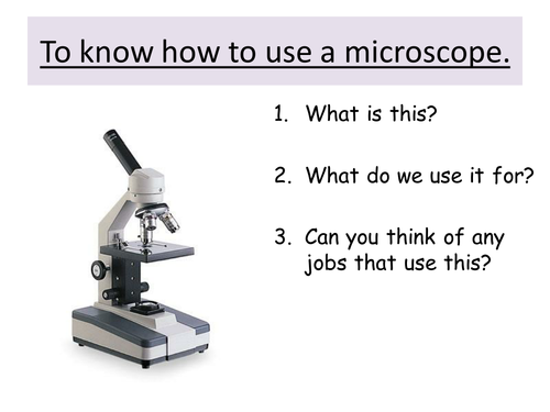
Microscope: Structure, Uses, Functioning Processes of Simple ...

Labeling the Parts of the Microscope | Microscope World Resources
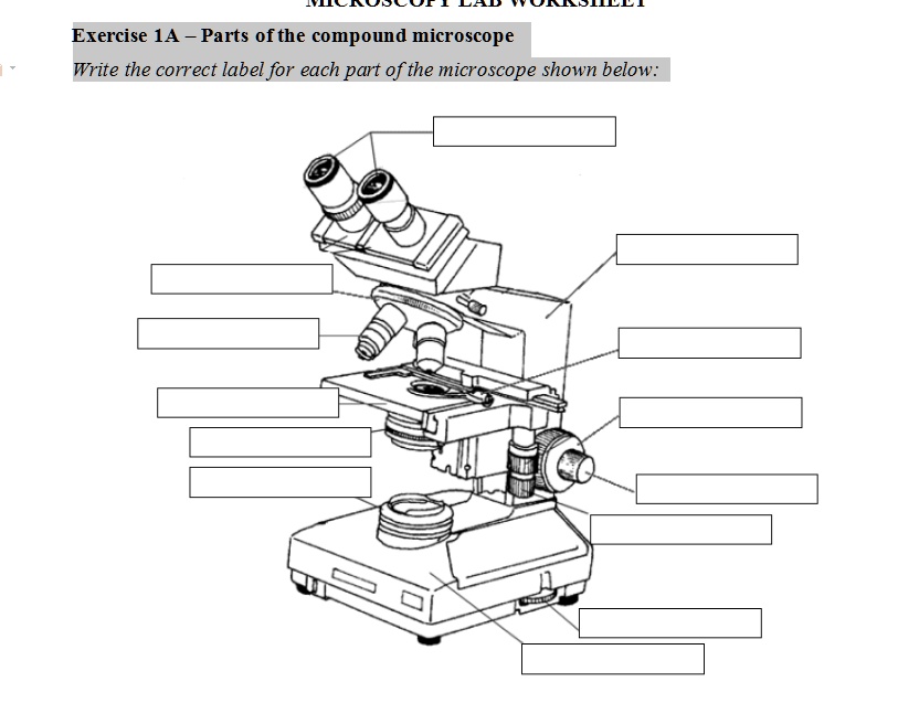
SOLVED: Exercise 1A Parts ofthe compound microscope Write the ...

Compound Microscope: Know Definition,working, diagram, properties

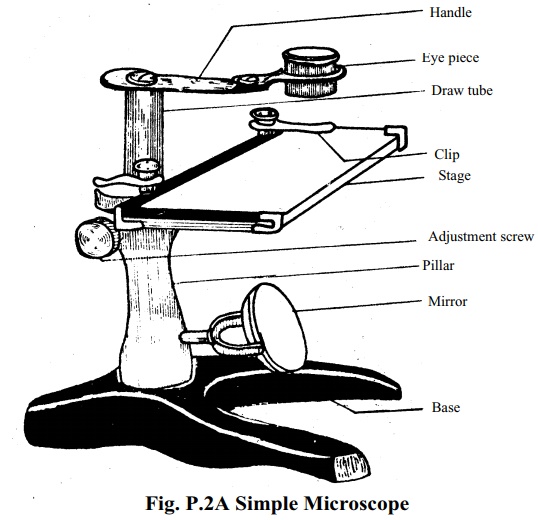
Simple Microscope - Diagram (Parts labelled), Principle ...

How to draw Microscope diagram for beginners - step by step

Microscope With Labels clip art | Microscope parts, Science ...
Label a microscope - Teaching resources

microscope drawing with label - Clip Art Library

Microscope labeled diagram
Glossary of terms used in microscopy – Quekett Microscopical Club

easy compound microscope diagram - Clip Art Library
What is Compound Microscope? - Diagram, Function, Advantages
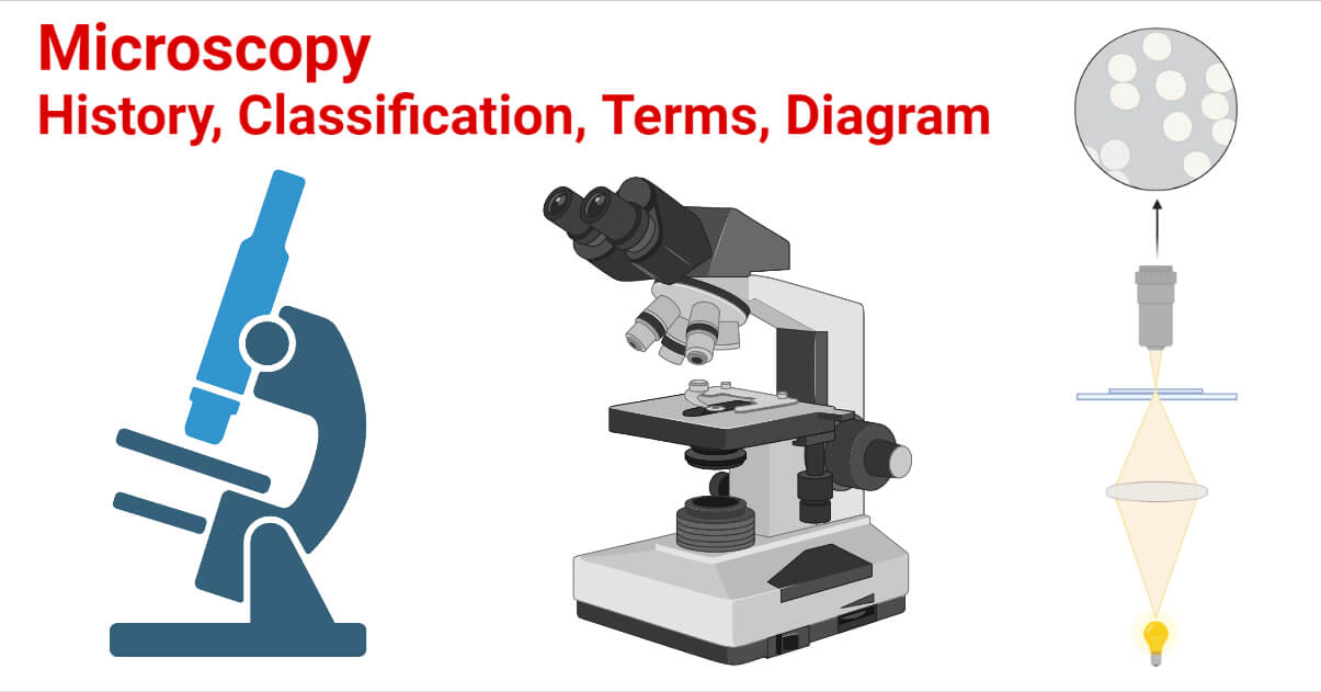
Microscopy- History, Classification, Terms, Diagram

How to draw compound of Microscope easily - step by step
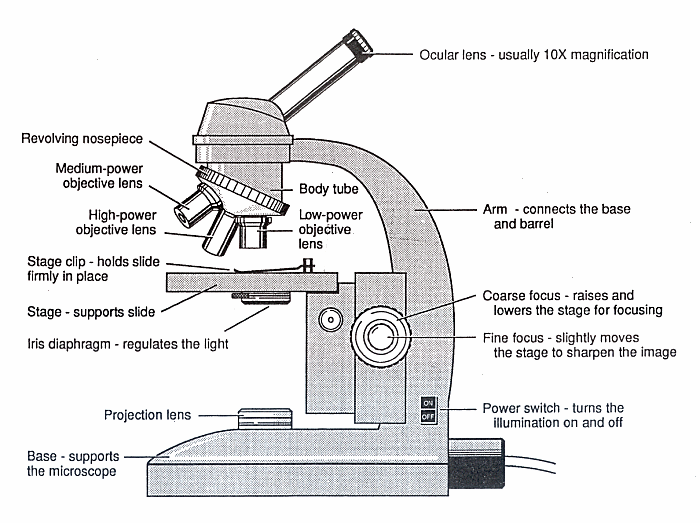
Diagram of a Microscope - Guide to using a microscope
PRACTICAL BOOKLET - BIOLOGY4ISC
Parts of a Microscope with Their Functions – Microbe Online

File:Labelledmicroscope.gif - Wikimedia Commons

Draw a neat labelled diagram of a compound microscope. Derive ...
Figure 1.15 A labelled diagram of a light microscope | Boost

Compound Microscope Parts – Labeled Diagram and their ...

Compound Microscope Parts, Functions, and Labeled Diagram ...

16 Basic Parts of Microscope, Function, Names & Labeled Diagram

Compound Microscope Parts – Labeled Diagram and their ...

Light Microscope- Definition, Principle, Types, Parts ...

Compound Microscope Parts, Functions, and Labeled Diagram ...

Draw a well labelled diagram of a microscope. - Brainly.in

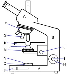
This is a common compound microscope. Label its parts from A ...

File:Microscope diagram.png - Wikipedia

Diagram of a Compound Microscope

Simple Microscope - Diagram (Parts labelled), Principle ...

draw a well label diagram of microscope - Brainly.in
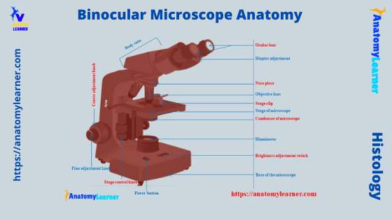
Binocular Microscope Anatomy - Parts and Functions with a ...

Microscope With Labels Clip Art at Clker.com - vector clip ...

Microscope Diagram Vector Illustration Labeled Zoom ...

Microscope diagram labeled | Clipart Panda - Free Clipart Images

Lable the microscope worksheet
Addgene: Using a Light Microscope Protocol

Dissecting Stereo Microscope Parts and Functions
Using a microscope | Teaching Resources


Komentar
Posting Komentar